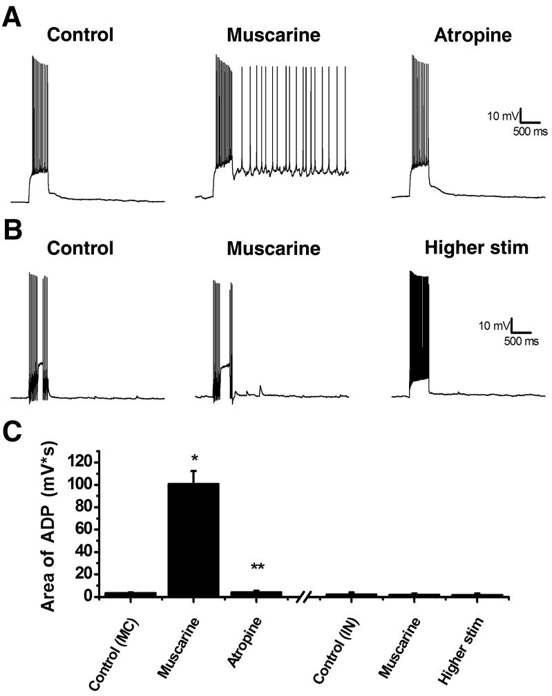
Figure 1. Muscarine can induce an ADP in mossy cells but not interneurons.
A, left: A mossy cell was held at a membrane potential of −60 mV and a 500 ms depolarizing pulse was applied to evoke a train of action potentials. After the depolarization the cell immediately returned to −60 mV. Middle: Following application of 5 μM muscarine an ADP was induced following the depolarizing current which could last for several seconds. Right: The ADP could be blocked by 5 μM atropine, a mAChR antagonist. B, left: A hilar interneuron was depolarized resulting in an immediate return to −60 mV. Middle: After application of muscarine, unlike mossy cells, there was no ADP. Right: An ADP could still not be produced with a larger somatic current injection. C: Summary plot showing the average area of ADP following the depolarizing pulse. The three bars left of the hash marks summarize the results of experiments in hilar mossy cells, while the three bars to the right of the hash marks are from hilar interneurons. *p<0.05 compared to baseline. **p<0.05 compared to muscarine. Higher stim was tested in 7 of 10 non-mossy hilar neurons.

