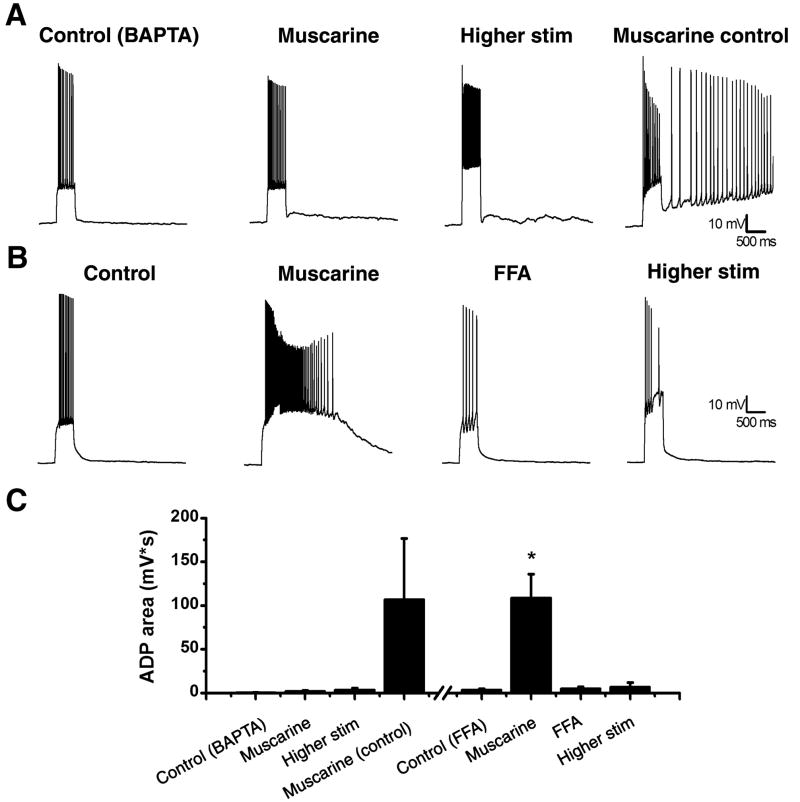
Figure 4. ADPs in mossy cells depend on calcium and depend on ICAN.
A: The internal solution was filled with 10 mM BAPTA, a calcium chelator, which blocked the ability to induce an ADP even with a higher stimulation (left, middle left, and middle right trace). A same day control showed an ADP could be produced with the muscarine (right trace). B: A representative cell before (left trace) and after (middle left trace) application of muscarine. The ICAN blocker, FFA (100 μM), blocked the ADP (middle right trace) and could not be recovered with higher stimulation (right trace). C: Summary plot of the ADP area under different conditions. For the BAPTA data set either 5 μM muscarine or 3 μM carbachol was used. *p<0.05 compared to control. **p<0.05 compared to muscarine.

