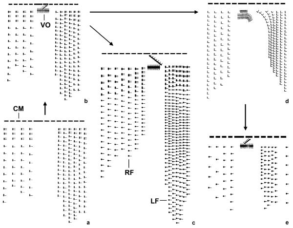
Fig. 2.
Kinetal maps showing the evolution of ciliary patterns in tintinnids with two ventral organelles. (a) The hypothetical ancestor of the tintinnids probably had a right and left ciliary field, and the anterior cilia of the dikinetids were reduced in the posterior portion of the kineties. (b) Two dikinetidal ventral organelles were introduced, resulting in the Tintinnidium (Tintinnidium) pattern (after Foissner and Wilbert 1979). (c) The bare anterior basal bodies of the dikinetids were partially lost in the posterior portion of the kineties, giving rise to the Tintinnidium (Semitintinnidium) pattern (after Blatterer and Foissner 1990). (d) The anterior cilia of the dikinetids were entirely lost, producing the Membranicola pattern (after Foissner et al. 1999). (e) The bare anterior basal bodies of the dikinetids were lost, creating the pattern of Tintinnopsis cylindrata (after Foissner and Wilbert 1979). CM, collar membranelles; LF, left ciliary field; RF, right ciliary field; VO, ventral organelles.

