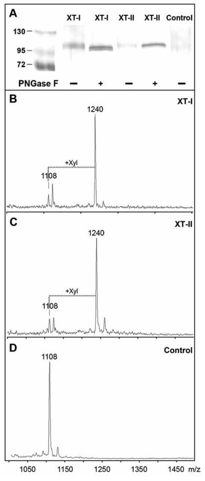
Figure 1. Expression of recombinant human xylosyltransferases in pgsA-745 cells.
(A) Western blotting of aliquots of culture supernatants of pgsA-745 cells transfected with either empty vector (Control), human XT-I or human XT-II were concentrated and, where indicated, subject to PNGase F digestion (+). Bands were detected using an anti-protein A antiserum as described. On the control blot, bands of Mr 26000 - 34000 were observed (region not shown) consistent with the expression of protein A alone in the control samples. Protein standards are indicated in kDa. (B-D) The culture supernatants were assayed overnight using a MALDI-TOF MS-based method. The syndecan peptide substrate (m/z 1108) was incubated in the presence of UDP-Xyl with culture supernatants of pgsA-745 cells transiently transfected with plasmids with either B) human XT-I, C) human XT-II or D) no insert. The increase in m/z of 132 is indicative of the transfer of xylose.

