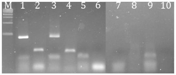
Figure 5. Presence of PCR inhibition after different stool RNA isolation techniques.

1% agarose gel electrophoresis of RT-PCR products shows inhibition of PCR by stool isolates using different purification techniques. M = DNA marker. Lanes 1 and 2 are the positive control using E. coli RNA with 16S rRNA gene primers primer or human leukocyte RNA with β-actin primer, respectively. Lanes 3 and 4 are the positive controls sequentially treated with bead-beating, RNA-Bee extraction, and silica column extraction, with 16S or β-actin primers, respectively. Lanes 5 and 6 are positive controls sequentially treated with lysis buffer homogenization and silica column extraction, with 16S or β-actin primers, respectively. Lanes 7 and 8 are positive controls sequentially treated with bead-beating and column extraction, with 16S or β-actin primers, respectively. Lanes 9 and 10 are positive controls sequentially treated with phenol:chloroform extraction and ethanol precipitation, with 16S or β-actin primers respectively.
