Figure 1.
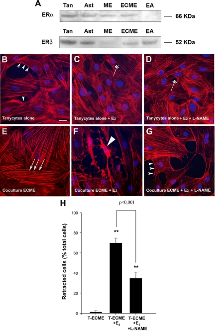
Ability of estradiol to influence endothelial cell-mediated plasticity in tanycytes. A, Western blot analysis of ERα and ERβ expression in primary cultures of tanycytes (Tan), cultured hypothalamic astrocytes (Ast), the median eminence (ME), immunopurified ECME, and the EAhy926 endothelial cell line (EA). Each well was loaded with 50 μg of protein. B–G, Tanycytes were cultured alone (B–D) or suspended above a conditioning layer of purified ECME (E–G) for 30 min and then stained with Alexa-conjugated phalloidin to visualize filamentous actin (red) and with Hoechst to stain nuclei (blue). Tanycytes alone (B–D) or both tanycytes and ECME (E–G) were cultured in the presence (C, D, F, and G) or absence (B and E) of 5 nm 17β-estradiol (E2) for 48 h (before coculture). Under basal conditions, when tanycytes were cultured alone (B), actin was localized adjacent to the cell membrane (cortical actin, arrowheads) and was also diffused throughout the cytoplasm. In contrast, tanycytes cultured with ECME for 30 min (E) exhibited bundles of actin filaments forming parallel stress fibers (arrows) running throughout the length of the cells but did not exhibit cortical actin. Note that although estradiol on its own had little effect on actin cytoskeleton remodeling in tanycytes (C), it promoted tanycyte retraction in coculture experiments (long arrowhead, F). Tanycyte retraction caused by endothelial cells in the presence of estradiol was prevented by pretreating endothelial cells with L-NAME (1 mm) (G), indicating that these plastic changes required endothelial NO secretion. *, Polymerized actin merged to puncta. Scale bar, 10 μm. H, Bar graph illustrating the percentage of tanycytes (T) that underwent retraction under various treatments. [ANOVA F(2,53) = 24.563, P < 0.001]. Error bars, sem. **, P < 0.001, as compared with untreated cocultures (T-ECME).

