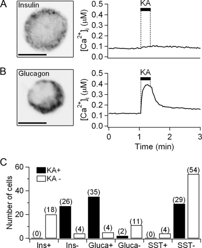
Figure 2.
Expression of iGluRs in immunocytochemically identified cells. A, An image of a single cell immunostained with antiinsulin antibody (scale bar, 5 μm) and the time course of Ca2+ response to 0.5 mmol/liter kainate (KA) for 20 sec. The cell was kainate unresponsive, relatively small, and insulin-positive. Insulin granules are labeled as punctate structures outside the nucleus. B, An image of another cell immunostained with antiglucagon antibody and its Ca2+ response to kainate. The cell was kainate responsive, small, and glucagon-positive. C, Summary bar graph showing the number of cells responding to kainate stimulation (KA+, black) or not responding (KA−, white). All the cells were sorted by their immunocytochemical identification as positive (+) or negative (−) for the indicated peptides. The numbers in parentheses indicated the total number of tested single cells.

