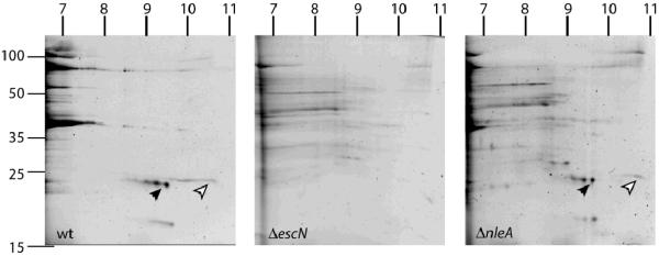
Figure 3. Map and EspF are secreted at wildtype levels by EPECΔnleA.
Sypro-stained 2-dimensional gels of secreted proteins from EPEC wildtype (left panel), EPEC ΔescN (center panel), and EPECΔnleA (right panel). Migration of molecular weight markers (in kiloDaltons) is indicated to the left of the first gel. Estimated migration of the pI gradients are indicated on the top of each gel. Map and EspF are indicated by the black and white arrowheads respectively. The identity of the proteins was verified by ion trap mass spectroscopy (see text).

