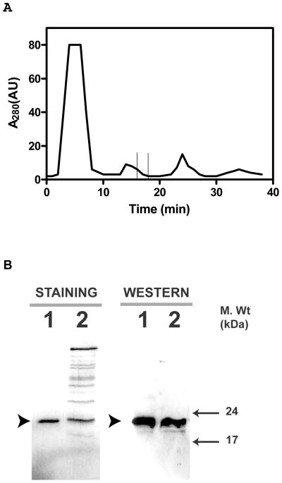
Figure 1. Purification of Hpca from bovine hippocampus.
Bovine Hpca was partially purified by hydrophobic interaction chromatography on phenyl-Sepharose CL-4B column as described in the “Materials and Methods” section. A. Elution Profile. The absorbance of the eluted proteins at 280nm was plotted against the elution time. The time at which Hpca elutes is marked by the window. B. Fractions containing Hpca were concentrated, separated by SDS-12%PAGE and analyzed by Coomassie blue staining and Western blotting. Band corresponding to Hpca is indicated by solid arrowhead. Lane 1. Recombinant myr-Hpca. Lane 2. Eluted and concentrated fraction.

