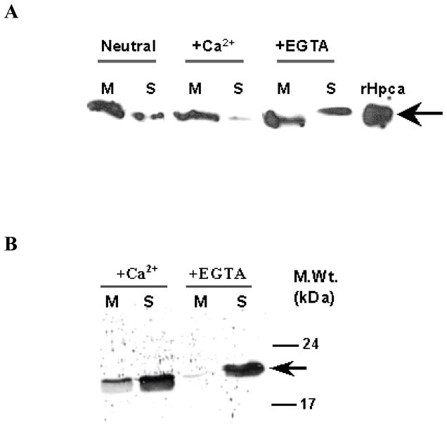
Figure 4. Calcium dependent distribution of Hpca in the membrane and soluble fractions of bovine hippocampus and COS cells.
(A) The membrane and soluble fractions of bovine hippocampus were prepared in the presence of 50 μM Ca2+ (+Ca2+), 2 mM EGTA (+EGTA) or in the absence of either (Neutral) as described in the “Materials and Methods” section. The membrane (M) and soluble (S) fractions (100 μg protein of each fraction) were individually separated by SDS-12%PAGE and subjected to Western blotting. 1 μg of recombinant Hpca was used as a positive control. (B) 5 μg of myristoylated Hpca was incubated with membrane fraction of COS cells (15 μg protein) in the presence of 50 μM Ca2+ (+Ca2+) or 1 mM EGTA (−Ca2+) as described in “Materials and Methods”. After 1 hr incubation the reaction mixture was centrifuged and the membrane (M) and soluble (S) fractions were analyzed by Western blotting using anti Hpca antibody. The position of the Hpca antibody immunoreactive band is indicated by an arrow. Molecular size markers are given alongside.

