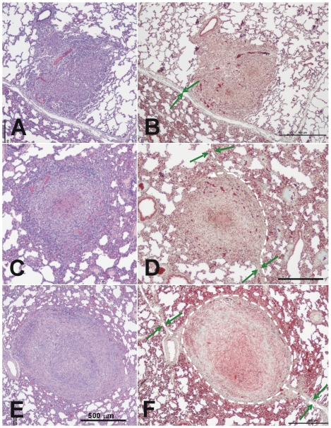
Figure 6. Microscopic evolution of recent lesions, showing the relationship between the granulomas and the intralobular septa.
A–D: Phase I lesions; E and F: Phase II lesions. Images A and B show the initial evolution phase where the granuloma touches an intralobular septa but there is still no fibroblast proliferation. This can be seen in images C and D, where the septa increase in thickness and start to surround the granuloma, as shown by the white dotted lines. Images E and F show how the granuloma is finally surrounded by a thick collagenic mantle. Pictures A, C and D were stained with haematoxylin and eosin (H&E), while B, D and F were stained with Masson's trichromic. The original magnification of the large images is ×40 whereas all insets, except F (x100), are magnified ×400. The green arrows show the septa and the dotted white lines the trajectory of the capsule.

