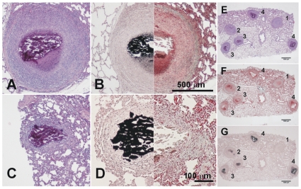
Figure 7. Microscopic evolution of the old lesions.
Once the granuloma is structured, the necrotic process starts and calcification appears. Images A and B (phase III) show a well-structured and encapsulated granuloma with necrotic calcification. Images C and D show lesions of an advanced evolution (phase IV), with granulomas containing a large amount of calcification and fibrosis. Samples A and C were stained with H&E, whereas samples B and D were half stained with Masson's trichromic and von Kossa stain, which shows calcification in black. Pictures E to G were stained with H&E, Masson's trichromic and von Kossa stain, respectively. Lesions 1 and 2 are phase II lesions and differ only in the initial mineralization seen in lesion 2. The other lesions are all phase III lesions that have progressed differently. Original magnification is ×10.

