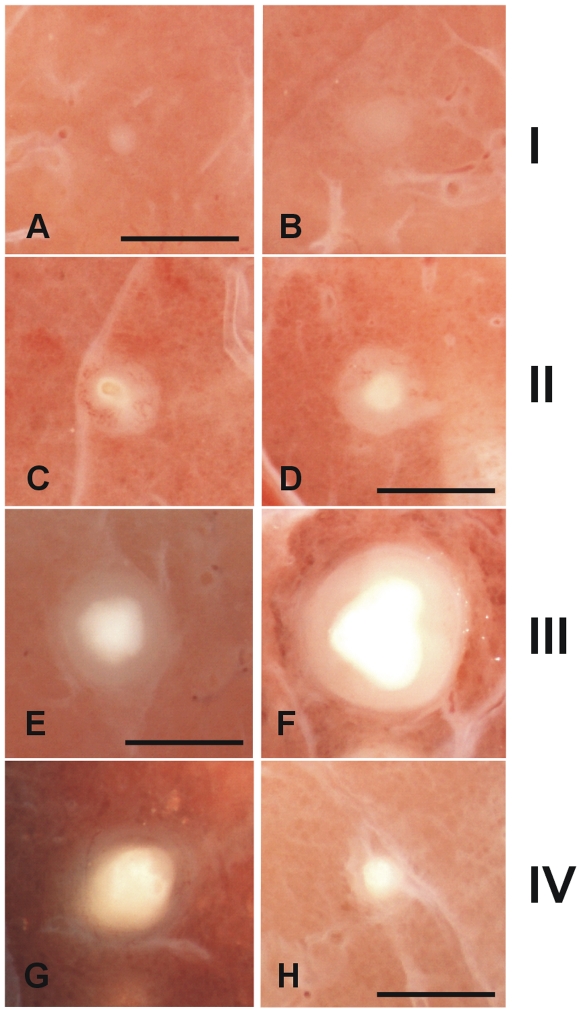
Figure 9. Macroscopic evolution of the lesions.
Classification of the fixed pulmonary lesions as they appear under the stereoscopic microscope. Considering the histological evolution of the lesions, and taking into account the sequential appearance of encapsulation and calcification, we have divided the lesions into four phases. Phase I is characterized by the presence of cellular infiltration (A and B). The intragranulomatous necrosis, which is characterized by the presence of an opaque zone inside the granuloma (C and D), appears during Phase II and structuration of the lesion starts. Phase III (E and F) involves the onset of calcification, which gives a shiny aspect to the central opacity, which grows in size. These lesions are characterized by the cartilaginous texture of the lesion when touched with the forceps. Phase IV lesions (G and H) are characterized by predominance of the calcification and a thin surrounding infiltration. Original magnification is ×10. Scale bar: 1 mm.

