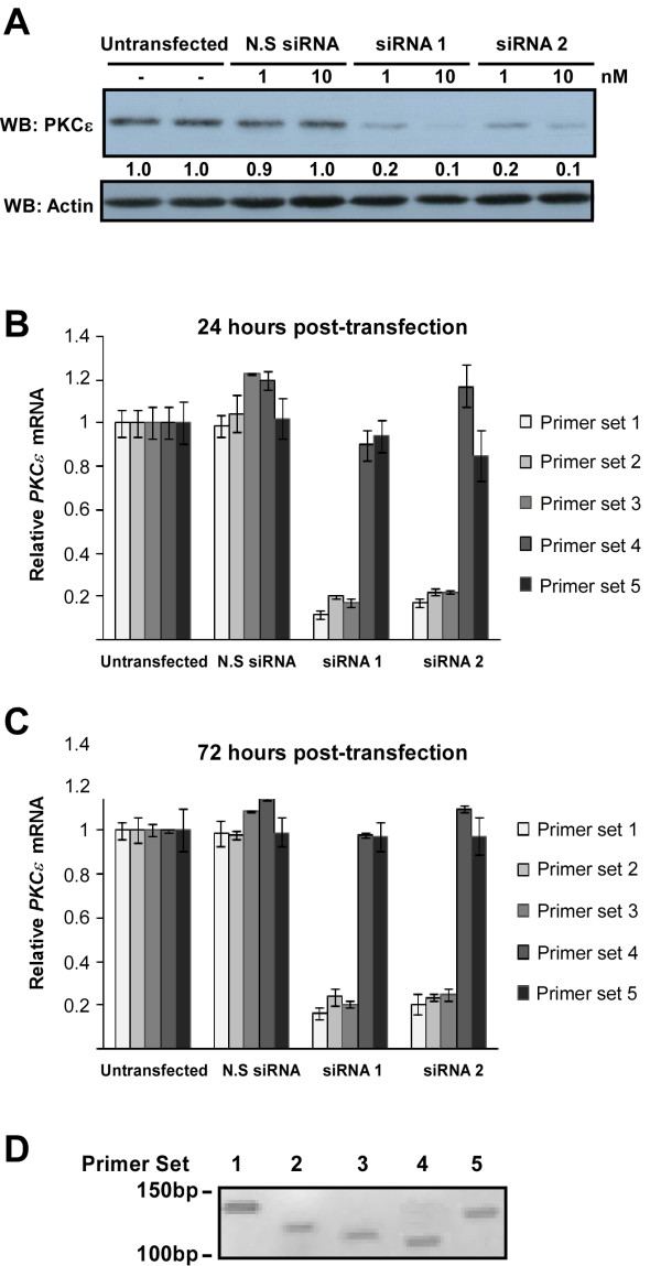
Figure 2.
Detection of PKCε knockdown by western blotting (WB) and RT-qPCR. HDMECs were treated with two siRNA duplexes to PKCε, or with a non-silencing control siRNA (N.S siRNA) at either 1 or 10 nM and RNA or protein prepared at 24-72 hours post-transfection. (A) Protein lysates prepared 48 hours post-transfection were analyzed for PKCε expression by western blotting (WB). Numbers below the blot denote the relative intensity of each band. The detection of actin expression was performed to monitor protein loading. (B) The amount of PKCε mRNA transcript at 24 hours post-transfection was analyzed by RT-qPCR using 5 different primer sets (typical Ct values for each primer set in untransfected cells were as follows: set 1: 25.7, set 2: 26.04, set 3: 25.8, set 4: 25.43, set 5: 25.51). Relative mRNA expression was determined using β-actin control (n = 3, mean ± S.D.). (C) The amount of PKCε mRNA transcript at 72 hours post-transfection was also assessed by RT-qPCR (typical Ct values: set 1: 25.82, set 2: 26.01, set 3: 25.92, set 4: 25.51, set 5: 25.64). Relative mRNA expression was determined using β-actin control (n = 3, mean ± S.D.). (D) Each primer set was tested to confirm a single PCR amplicon, visualized by agarose gel electrophoresis.

