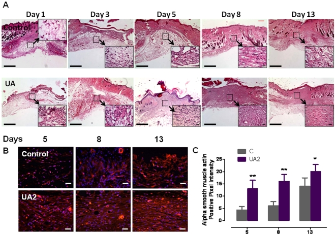
Figure 4. Histological features of wound healing in mice treated with UA2 or vehicle.
A. Images of skin tissue sections stained with hematoxylin and eosin showing histological changes during the wound healing process in control mice, with uric acid analog at post-injury days 1, 3, 5, 8 and 13. UA2 treated mice exhibited enhanced restoration of dermal and epidermal tissues in the wound. See File S1 for a detailed description of histological changes in the different groups of mice. Scale bar = 1 mm. These images are representative of 12 wounds in 6 mice for each treatment group. B. Immunostaining for α smooth muscle actin showing wound healing tissues on days 5, 8 and 13 in mice treated with UA2 or vehicle. Pictures showing enhanced myofibroblast differentiation in UA2 treated groups compared to controls in days 5, 8 (**p<0.01) and 13 ((*p<0.05). Scale bar = 25 µm. C. Quantification of alpha smooth muscle actin positive staining.

