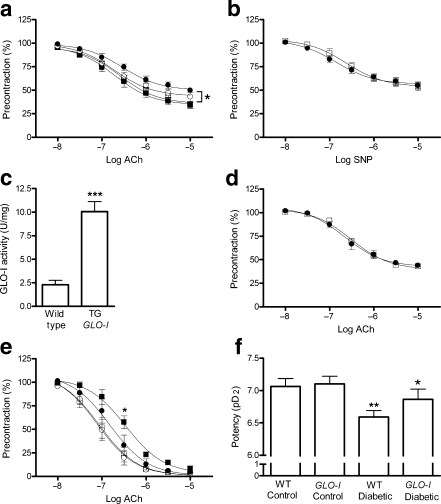
Fig. 1.
Effect of high glucose concentration on acetylcholine (ACh)-induced NO-mediated endothelium-dependent relaxation in isolated rat mesenteric arteries. a Rat mesenteric arteries were mounted in a myograph and incubated with or without high glucose concentrations for 2 h. Acetylcholine-induced NO-mediated endothelium-dependent relaxation was impaired in a concentration-dependent manner during incubation with 30 mmol/l glucose (white circles) or 40 mmol/l glucose (black circles) compared with control (5 mmol/l glucose, white squares). The 40 mmol/l glucose incubation resulted in a statistically significant shift of the dose–response curve (*p < 0.05 vs control group). Incubation with 35 mmol/l mannitol (black squares) as an osmotic control did not impair NO-mediated endothelium-dependent relaxation (n = 6). b SNP-induced NO-mediated endothelium-independent vasorelaxation during incubation without (white squares) or with 40 mmol/l glucose (black circles) for 2 h did not differ (n = 6). c GLO-I activity in mesenteric arteries homogenates of GLO-I transgenic (TG) rats was significantly elevated compared with mesenteric arteries homogenates of wild-type rats (***p < 0.001, n = 4). d In mesenteric arteries of GLO-I transgenic rats, acetylcholine-induced NO-mediated endothelium-dependent relaxation was not impaired during incubation with 40 mmol/l glucose (black circles) for 2 h compared with control incubations (5 mmol/l glucose, white squares, n = 5). e Acetylcholine-induced vasorelaxation was significantly impaired in wild-type diabetic rats (black squares; *p < 0.05) compared with the controls (wild-type control, white squares; transgenic control, white circles). GLO-I overexpression improved this relaxation (black circles; n = 8). f The decreased potency in wild-type (WT) diabetic rats (**p < 0.01) was significantly improved by GLO-I overexpression (*p < 0.05)

