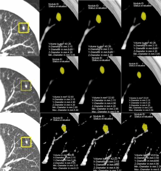
Fig. 2.
Volume-rendered display showing three repeated measurements of a sub-centimetre intra-parenchymal pulmonary nodule with reconstruction settings A (top), B (middle) and C (bottom). Nodule measurements are completely identical with setting A (40.3 mm3), slightly different with setting B (32.9, 32.9, 33.0 mm3) and moderately different with setting C (39.1, 42.2, 41.1 mm3). The means of measured volume with settings A and C were larger than with setting B (40.3 vs 32.9 vs 40.8 mm3)

