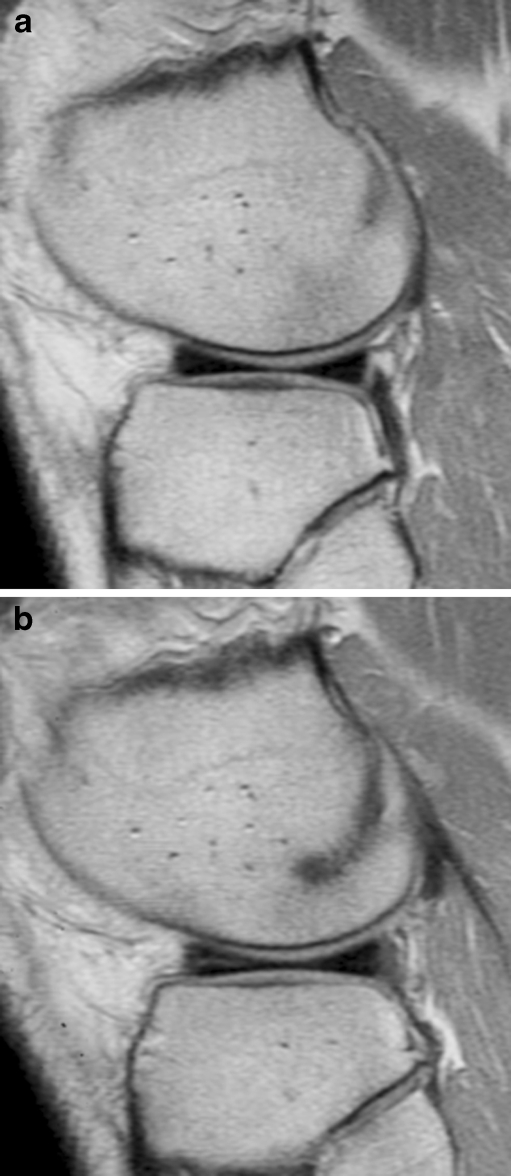
Fig. 2.
Development of a degenerative lesion on follow-up MRI in a patient who sustained a distortion of his right knee. a On the initial MRI examination, a normal anterior horn of the lateral meniscus was seen. b Follow-up MRI after 1 year demonstrated a linear band of increased signal intensity that did not extend to the articular surface, which was scored as a grade 2 degenerative lesion

