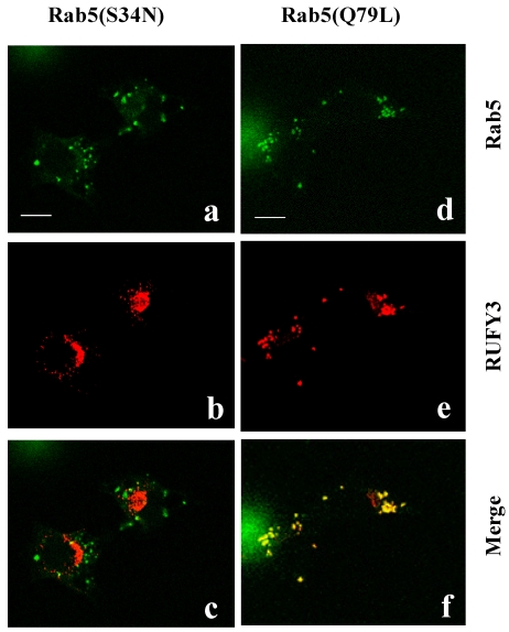
Figure 1.
Colocalization of RUFY3 and active Rab5(Q79L). The 3Y1 cells were transiently transfected with either GFP-tagged-Rab5(S34N) and pcDNA3.1-RUFY3 (left panels), or GFP-tagged-Rab5(Q79L) and pcDNA3.1-RUFY3 (right panels). The cells were then fixed and incubated with anti- RUFY3 antibodies followed by staining with rhodamine-conjugated secondary antibodies. Images were obtained using a confocal microscope. The merged figures show images of green (GFP-labeled proteins) and red rhodamine labeling obtained for the same section, with yellow color resulting from the overlay of green and red. Polyclonal anti-RUFY3 antibody was prepared in rabbits immunized with the NH2-terminal region of human RUFY3 as GST fusion proteins. Antibodies (Rab5:sc46692; Rab5A:sc309) were purchased from Santa Cruz Biotechnology. Secondary antibodies linked to Fluorescein or Rhodamine were from BIO SOURCE. Bars, 10 micrometer.

