Figure 2.
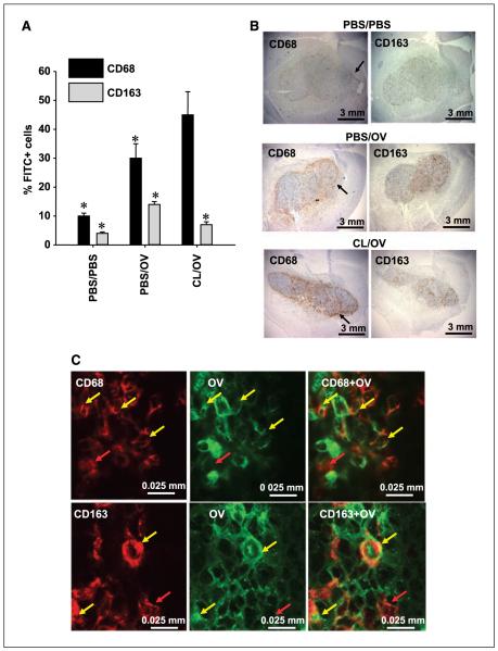
CL-mediated depletion of tumor-associated phagocytic cells. A, FACS analysis of the brain hemispheres harboring gliomas from rats treated with mock solution (PBS/PBS), OV alone (PBS/OV), or CL (CL/OV) before OV delivery. The graph shows the average percentage of CD68+ and CD163+ cells, detected by FACS, within the whole brain hemisphere of each treatment group (n = 3). For each cell type, a 3-fold increase is observed following OV treatment, and this increase is inhibited by CL treatment only for CD163+ cells. Significance of pair means comparison was determined by ANOVA followed by post hoc Tukey’s test for each cell type (CD68: P = 0.0085; CD163: P = 0.011). Asterisks, significant differences. No significant difference was observed for CD68+ cells in animals treated with PBS/OV and CL/OV. B, microphotographs of brains with established D74/HveC syngeneic gliomas for rats treated with mock vehicle (PBS/PBS, top row), OV alone (PBS/OV, middle row), and CL (CL/OV, bottom row) before OV injection. CL-mediated macrophage depletion is observed only for the CD163 antigen (right column), whose intratumoral distribution varies somewhat from that of the CD68+ cells (left column). Black arrows, accumulation of CD68+ cells around the tumor edge. C, microphotographs of OV-infected cancer cells in a rat tumor treated with OV for 72 h. The brains were stained with antibodies recognizing HSV1 antigens (green) and either anti-CD68 or anti-CD163 antibodies (red). Clones of OV-infected cancer cells are surrounded by CD68+ and CD163+ cells. Some of these are localized in the proximity of HSV and remain red (red arrows), but others have clearly incorporated HSV-infected cells, and colocalization of the two markers gives an orange-yellow staining (yellow arrows). The incorporation of HSV1 into either CD68+ or CD163+ cells was confirmed by scanning the laser through the slide with 1-μm intervals.

