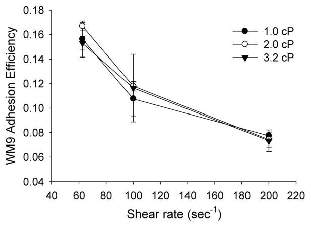
Figure 3. Effects of shear stress and shear rate on melanoma adhesion to the EC in the presence of PMNs.
The parallel plate flow chamber was perfused with appropriate medium over the EI monolayer for 2–3 minutes at a shear rate of 40 sec−1 for equilibration and then PMNs and WM9 cells were injected into the flow chamber through the syringe pump at a predetermined concentration (1×10⁶ cells/ml) of each. The cell-cell interactions in shear flow were then recorded and analyzed off-line. WM9 adhesion efficiency was defined in “Materials and Methods”. Values are mean ± S.E.M. for N ≥ 3.

