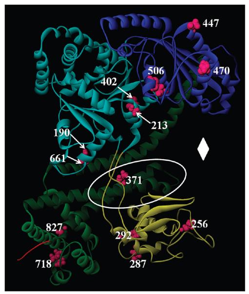
Figure 1. Location of SecA domains and cysteine residues.
A model of the NMR structure of E. coli SecA (Protein Data Bank entry 2VDA structure 1) (38) is shown in ribbon representation. The structure of SecA is colored according to domains: light blue for NBD-1, dark blue for NBD-2, yellow for PPXD, dark green for HSD, light green for HWD, and red for the structured portion of CTL. The SecA monocysteine residues used in this study are colored magenta and are labeled with their residue number. The two-helix finger is in the region that falls within the white oval, and the clamp region is identified with a white diamond (10).

