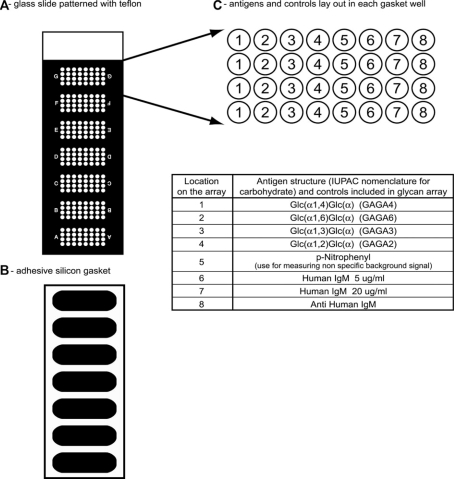
Figure 1.
Glycan array format: A – Glass slide patterned with Teflon mask creating 7 clusters of microwells, 32 wells in each cluster. B – An adhesive silicon superstructure attached to the slide defines wells for manual application of multiple serum samples per slide. C – Antigens and controls lay out in each gasket well.

