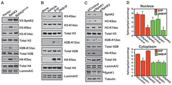
Fig. 2.
SphK2 and S1P enhance histone acetylation. (A) Histone acetylation in nuclear extracts from MCF-7 cells transfected with vector, V5-SphK2, or catalytically inactive V5-SphK2G212E. Acetylation was detected by immunoblotting with antibodies to specific histone acetylation sites as indicated. (B) Effect of S1P and dihydro-S1P on histone acetylation. Purified nuclei from MCF-7 cells were treated for 5 min without (none) or with vehicle, 1 μM S1P, or 1 μM dihydro-S1P, and histone acetylation was examined by Western blotting. (C and D) Effect of depletion of SphKs. MCF-7 cells were transfected with nontargeting control siRNA, or siRNA targeted to SphK1 or SphK2. (C) Proteins from nuclear extracts were immunoblotted as indicated. Depletion of SphK1 and SphK2 was confirmed by immunoblotting with SphK1-specific and SphK2-specific antibodies, respectively. Asterisk (*) indicates nonspecific band. (D) Amounts of S1P and dihydro-S1P in purified nuclei and cytoplasm quantified by LC-ESI-MS/MS. Asterisks indicate statistically significant differences (P < 0.05 by Student’s t test) relative to control siRNA.

