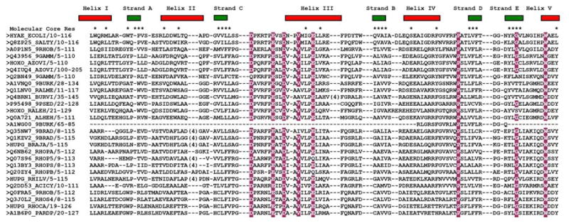
Fig 2.
Multiple sequence alignment of the members of Pfam family PF07449. The sequences of HYAE_ECOLI and Q8ZP25_SALTY are shown in the top two rows. The secondary structure elements of Q8ZP25_SALTY are shown above the alignment, and residues participating in the molecular core are marked with an asterisks (*). Conserved residues with 85% or greater conservation are highlighted in burgundy. Those residues are also seen in the most conserved group resulting from the analysis using the program Consurf analysis (Figure 3a). For comparison, the sequence of thioredoxin (PDB ID 1THX) was aligned with the sequence of protein Q8ZP25_SALTY using a structure based alignment algorithm [40] and is shown at the bottom. The Cys-X-X-Cys motif characteristic of thioredoxins is highlighted in blue (and absent in PF07449).

