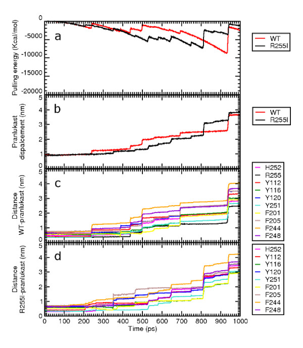
Figure 8.
Forced unbinding profile of pranlukast. Panel a and b compare the unbinding simulations of pranlukast from the WT (in red) and the R255I (in black) receptor models: panel a shows the work developed to unbind pranlukast; panel b shows the displacement of the COM of the ligand from its starting position. Panel c and d show the distances between groups of atoms of the ligand that form polar or hydrophobic interactions with atoms of the WT or the R255I models, respectively.

