
Fig. 6.
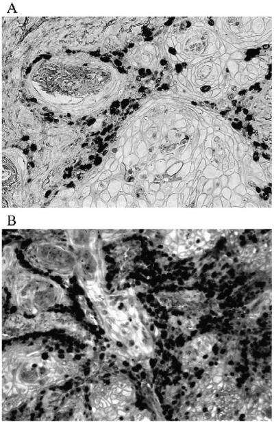
Ki-67 staining of tumor tissue from UROtsa (A) and URO-MSC52 (B) cells. Representative photograph of Ki-67-stained tissue sections demonstrates the increased proliferation found in URO-MSC52 tumors. Magnification used for photographs is ×400.