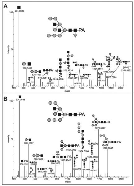
Figure 3. MS/MS analysis of the major N-glycans of axenically-grown mid-log cells.

The major AX3 pyridylaminated N-glycans were purified by sequential NP-HPLC (Palpak) and RP-HPLC prior to analysis by LC-ESI-MS/MS; the MaxEnt3 transformed data for (A) the m/z 1176.93 [M+2H]2+ (Man8GlcNAc4Fuc1) and (B) the m/z 1103.41 [M+2H]2+ (Man8GlcNAc4) molecular ions are shown. Fragments of the postulated complete glycan structures are depicted using the nomenclature of the Consortium for Functional Glycomics (black square, N- acetylglucosamine; grey circle, mannose; grey triangle, fucose). The composition and linkages of the intact glycans were proven by GLC-MS (see Table 1); up to three mannose and one GlcNAc residues could be released by combined α-mannosidase and β-N-acetylhexosaminidase digestion (data not shown). Based on the expected prior processing by ER mannosidase, the Man8B isomer is taken as the basis for these structures.
