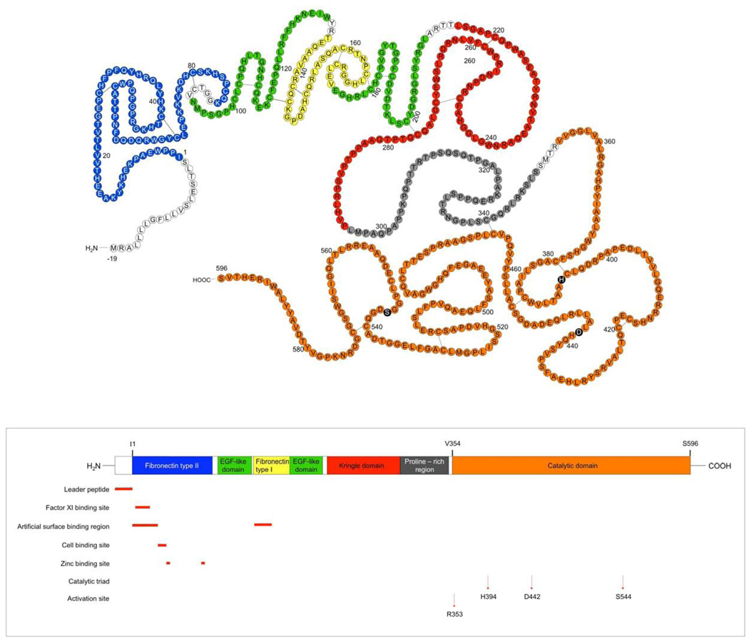
Figure 1.
Structural Basis of FXII Function: FXII is divided into several domains. Top structure: amino acid sequence; bottom structure, linear diagram color coding each of the regions on the protein. Amino acids -19-1: leader peptide, 1–88: fibronectin type II domain, 94–131: EGF-like domain, 133–173: fibronectin type I domain, 174–210: EGF-like domain, 215–295: kringle domain, 296–349: proline-rich region, 354–596: catalytic domain or light chain. Amino acids 1–353 are the so-called heavy chain. Each of these areas is highlighted in the same color as the linear cartoon below it. This figure is adapted from Cool and MacGillivray [5].

