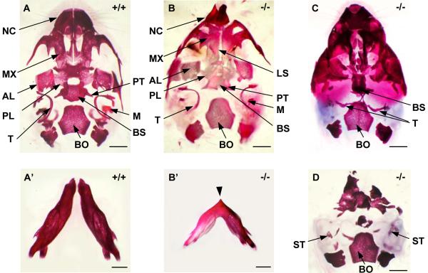
Fig. 3. Ventral views of the whole-mount skull stained with alizarin red.
(A) Wild-type ventral skull with normal mandibular bones (A') at birth. (B-D) Twsg1−/− skulls; (B) Ventral skull of Twsg1−/− newborn mouse with micrognathia showing deformity of the nasal capsule (NC), a longitudinal slit (LS) in the skull base, and preservation of the malleus (M) and pterygoid (PT). (B') Mandible in a micrognathic Twsg1−/− newborn mouse; the mandibular bones are fused in the midline indicated by an arrow. (C) Midline fusion of the right and left tympanic bones (T) in the agnathic Twsg1−/− mouse. (D) Loss or deformity of the majority of skeletal elements anterior to basioccipital (BO) with preservation of stapes (ST) in the Twsg1−/− mouse with anterior truncation phenotype. AL, alisphenoid; BS, basisphenoid; MX, maxilla; PL, palatine. Scale bar: 1 mm.

