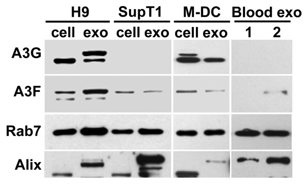
Fig. 2. Expression of A3G and A3F in exosomes.
Exosomes were purified from cell culture supernatants or from blood plasma of 2 different healthy blood donors. Cell and exosome lysates (20 and 10 µg/lane, respectively) were separated by SDS-PAGE and analyzed by Western blotting for the presence of A3G, A3F, and exosome markers Rab7 and Alix. In addition to the 70–75 kDa form of Alix detected predominantly in cells, higher molecular forms of Alix (95–105 kDa) were detected, particularly in exosomes. A slower migrating form of A3G was specifically detected in H9 exosomes. Note that A3F was detected in exosomes purified from blood plasma of donor 2 but not donor 1.

