
Figure 1. CXCR4 protein expression in colorectal cells was analyzed by immunocytochemistry.
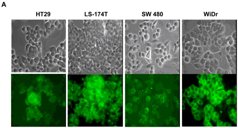
A. Colorectal cancer cell lines were evaluated for CXCR4 receptor expression. The top row is unstained cells and the bottom is IHC staining for CXCR4 using flourescein isothiocynatae (FITC) green. B. Breast cancer cells, MDA-MB-231 and MDA-MB- 468 were positive and negative controls, respectively. Magnification: 400x.