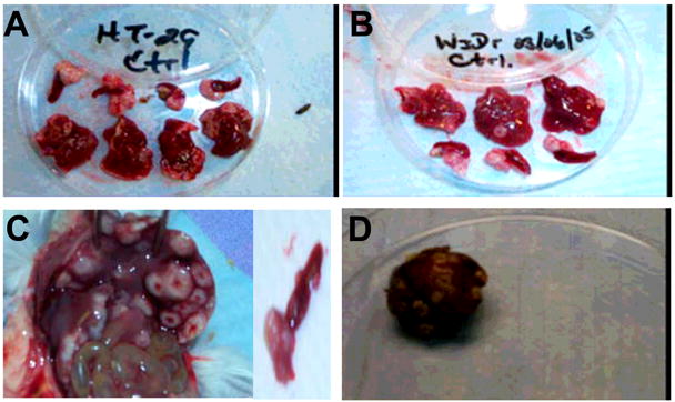
Figure 3. Primary tumor growth and liver metastasis.

Spleens and livers excised from the SCID mice four weeks after spleens were inoculated with tumor cells (A-HT-29, B-WiDr, C-LS174t, D-SW480). Primary tumor growth was seen in the spleens of all mice. All mice were untreated. Pictures were taken using a Kodak DC290 zoom digital camera.
