
Figure 1. Visualizing aromatase expressing neurons in the mouse brain.
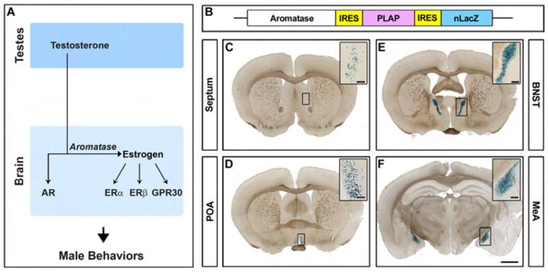
(A) Sex steroid hormone control of male behaviors. (B) Schematic of genetic modification of the aromatase locus. (C–F) Coronal sections through the forebrain of an adult male homozygous for the aromatase-IPIN allele stained for βgal activity. Aromatase is expressed in a sparse manner in discrete regions including the lateral septum (C), preoptic area (POA) (D), bed nucleus of the stria terminalis (BNST) (E), and medial amygdala (MeA) (F). Scale bar equals 2.5 mm. Inset scale bars equal 200 μm (C, D) and 50 μm (E, F).