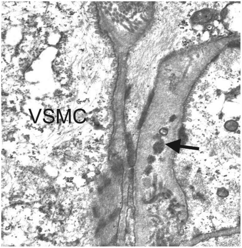
Fig. 1.
Characteristic pathologic findings in CADASIL on electron microscopy. An arteriole obtained during a skin biopsy shows multiple GOMs (arrow) between the basement membranes of VSMC (Courtesy of Sung-Hye Park, MD, PhD). CADASIL: cerebral autosomal dominant arteriopathy with subcortical infarcts and leukoencephalopathy, GOMs: granular osmiophilic materials, VSMC: vascular smooth-muscle cell.

