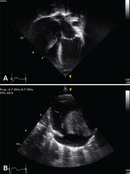
Figure 2).
A Apical four-chamber two-dimensional echocardiographic image demonstrating a large myxoma arising from the anterolateral atrial wall obstructing right ventricular inflow. B Modified parasternal long-axis two-dimensional echocardiographic view demonstrating the myxoma obstructing the tricuspid valve and right ventricular inflow

