Figure 3.
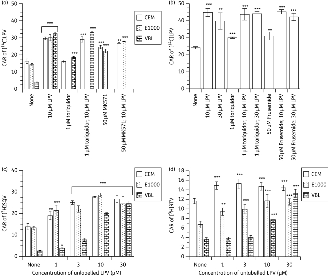
Effects of (a) pre-incubating CEM, CEME1000 (E1000) and CEMVBL (VBL) cells with fixed concentrations (30 µM) of unlabelled lopinavir (LPV) (alone and in combination with 1 µM tariquidar or 50 µM MK571), (b) co-incubating the PBMCs with 10 and 30 µM unlabelled LPV (alone and in combination with 1 µM tariquidar or 50 µM frusemide) on the accumulation of [14C]LPV, (c) pre-incubating CEM, E1000 and VBL cells with various concentrations (0–30 µM) of unlabelled LPV followed by the addition of [3H]saquinavir (SQV) and (d) pre-incubating CEM, VBL and E1000 cells with various concentrations (0–30 µM) of unlabelled LPV on the accumulation of [3H] ritonavir (RTV). Bars indicate mean ± SD (n = 4, with four independent observations from cultured CEM and its variant cells and n = 4 with four independent observations from each buffy coat PBMC sample). P values of *P < 0.05, **P < 0.01 and ***P < 0.001 indicate statistically significant differences in the CAR of [14C]LPV or [3H]SQV between controls and drug-treated samples. Note that tariquidar/LPV combinations were compared with samples treated with tariquidar alone.

