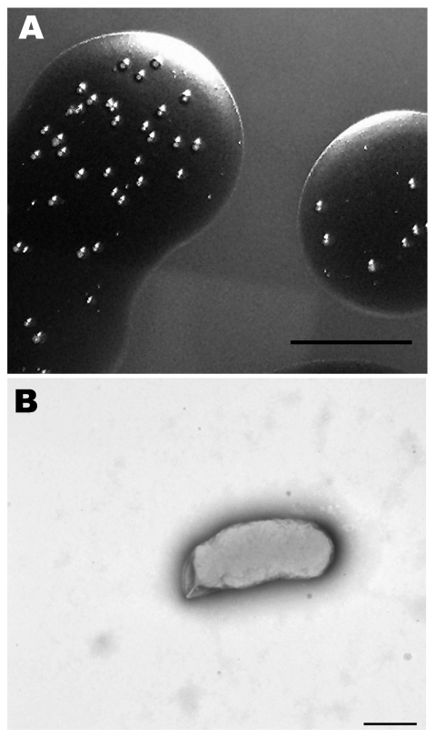
Figure 1.
Morphologic analysis of a Bartonella sp. isolated from sheep blood. A) Colonies growing in sheep blood surface biofilm seen in reflected light after 25 days. Scale bar = 10 mm. B) Transmission electron micrograph of a representative cell that was dispersed from a 25-day-old colony and negatively stained with 0.5% potassium phosphotungstic acid. Scale bar = 500 nm

