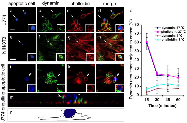
Figure 5. Dynamin in phagocytes is recruited around apoptotic cells coincident with corpse internalization.
CFSE/TAMRA stains the cytoplasm of apoptotic cells, frequently resulting in a lunette of staining around the nucleus. Arrows and asterisks indicate apoptotic cells. Scale bar, 10 Îm. Error bars represent s.d.
(aâh) J774 macrophages (aâd) or NIH/3T3 fibroblasts (eâh) were incubated with apoptotic thymocytes or apoptotic Jurkat cells, respectively, and localization of endogenous dynamin (green) and polymerized actin (red) were monitored. Dynamin was recruited to the phagocytic cup with actin in J774 (15 min) (d, merge) and NIH/3T3 cells (30 min) (h, merge) (indicated by arrow), but was not recruited around bound apoptotic cells (d, arrowhead).
(iân) Confocal z-sections were reconstructed to generate yz planes (m, n). J774 macrophages incubated with apoptotic cells at 4 ÂC did not show phagocytic cup formation or enrichment of endogenous dynamin around the apoptotic cell (Supplementary Figure S6). Cells incubated at 37 ÂC showed dynamin localized in the phagocytic cup (m, arrowhead and n, camera lucida) adjacent to the apoptotic cell in a punctate pattern (j, k, inset). Dotted lines (l) indicate plane of yz reconstruction.
(o) Time course of endogenous dynamin recruitment around the apoptotic cell corpse in J774 macrophages. Cells that had bound apoptotic cells were scored positive for dynamin recruitment if a âhaloâ of dynamin was seen to surround the apoptotic cell. 4 ÂC, n=100 (4 experiments); 37 ÂC, n=200 (8 experiments).

