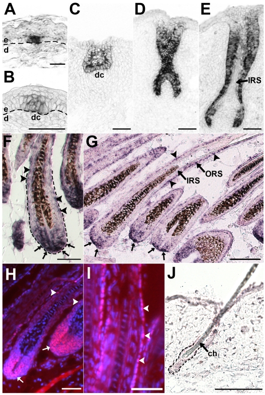
Figure 1. Lhx2 is expressed from early stages of morphogenesis and becomes restricted to the proximal part of the hair bulb and the ORS in fully developed HFs.
Lhx2 expression analysed by in situ hybridization (A–G,J) and immunohistochemistry (H-I) of tissue sections of HFs at different stages of morphogenesis (A–F) and in postnatal anagen (G–I) and telogen (J). (A) Lhx2 expression in basal keratinocytes at the pre-germ stage (Stage 0) prior to any obvious morphological change of the keratinocytes or the underlying dermis. (B) HF at the hair germ or placode stage (Stage 1) showing Lhx2 expression in the keratinocytes located at the local thickening of the epidermis. Reorganisation of mesenchymal cells in the dermis beneath the placode is indicative of formation of a dermal condensate. (C) HF at germ/peg stage (Stage 2–3) of HF morphogenesis. (D) HF at the peg stage (Stage 4), Lhx2 is expressed in the entire epithelial portion of the HF. (E) HF at the bulbous peg stage (Stage 5–6) when formation of the IRS has begun. Lhx2 is still widely expressed in outer layers of the HF and in the proximal extension the ORS in the future hair bulb whereas expression is turned off in cells in the forming IRS. (F) Fully developed HFs when the hair shaft has erupted through the epidermis (Stage 8). Lhx2 is expressed in the proximal part of the hair bulb (arrows) and in cells scattered in the ORS (arrow heads). (G) Analysis of Lhx2 expression in HFs during postnatal anagen Sub-stage VI revealing expression in the proximal part of the hair bulb (arrows) and in cells scattered in the ORS (arrow heads). (H,I) Immunohistochemical analysis of Lhx2 expression in anagen Sub-stage VI HFs revealing presence of nuclear Lhx2 protein in cells in proximal part of the hair bulb and in matrix cells (H, arrows) and in cells scattered in the ORS (H and I, arrow heads). (J) Analysis of Lhx2 expression in adult HFs in the extended telogen in 7–8 weeks old mice revealing no detectable expression. ch, club hair; dc. dermal condensate; d, dermis; e, epidermis. Scale bars, (A–I) 50 µm; (J) 100 µm.

