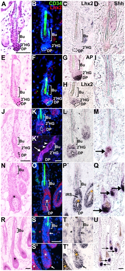
Figure 2. Lhx2 expression is associated with anagen during postnatal HF cycling and is not transcribed by CD34+ bulge cells.
HFs at different Stages and Sub-stages of postnatal cycles analysed by Hematoxylin/Eosin (H/E) staining (A,E,J,N,R), CD34 immunofluorescence (B,F,K,O,S,S’), Lhx2 in situ hybridisation (B,C,F,H,K,L,O,P,S,S’,T,T’), Lhx2 immunofluorenscence (K’), Shh in situ hybridisation (D,I,M,Q,U) and alkaline phosphatase (AP) staining (G). The CD34 immunoflouresence and Lhx2 in situ hybridisation are performed on the same section and the Lhx2 in situ signal (black) has been pseudocoloured red on the DAPI stained sections (B,F,K,O,S,S’). AP staining distinguishes the DP from the secondary HG where Lhx2 is initially expressed in late telogen (F–H). Shh expression confirms the stage of the HF cycle since Shh is only expressed during anagen. (A–D) HFs in early telogen, CD34+ cells are located in the bulge (Bu) area (B) and no Lhx2 expression (B and C) or Shh expression (D) can be detected at this stage. (E–I) HFs in late telogen, CD34+ cells are located in the bulge area (F) whereas Lhx2 is expressed by cells in the secondary hair germ (2°HG) located between the bulge area and the AP+ cells in the DP (F–H). There is no overlap in Lhx2 and CD34 expression (F). Shh is not expressed confirming that the HFs are in telogen (I). (J–M) HFs in anagen Sub-stages I-II as no deposition of pigment is detected (J). CD34+ cells are located in the bulge area whereas Lhx2 is expressed in the secondary HG and the extended part of the HF enveloping the DP (K and L). Immunohistochemical analysis of Lhx2 protein reveal presence of Lhx2 in the 2°HG (K’, arrows) as well as a few cells in the lower part of the bulge region (K’, arrow heads), despite absence of Lhx2 mRNA in this part of the HF. Shh is expressed confirming that anagen has commenced (M, arrow). (N–Q) HFs in anagen Sub-stage III as deposition of pigment has started (N). CD34+ cells are located in the bulge area (O) whereas Lhx2 expression is detected in the lower transient part of the HF (O,P). Shh is expressed confirming ongoing anagen (Q, arrows). (R–U) HFs in anagen Sub-stage IV-V as the hair shaft has reached the hair canal (R). CD34+ cells are located in the bulge area whereas Lhx2 expression is detected in the proximal part of the hair bulb (S’ and T’, arrow) and in cells scattered in the ORS (S and T, arrow). Shh is expressed confirming ongoing anagen (U, arrows). * indicates melanin deposition. Scale bars, 50 µm. 2°HG, secondary hair germ; Bu, bulge; DP, dermal papilla; IRS, inner root sheath.

