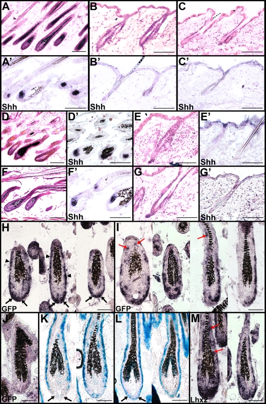
Figure 5. Lhx2 expression is sufficient to induce the anagen stage of the HF cycle.
(A,A’) H/E staining of back skin HFs in anagen in 9 weeks old Tx-treated Z/Lhx2-GFP:CreER mice (A) revealing Shh expression (A’). (B,B’) H/E staining of back skin HFs in telogen in 9 weeks old Tx-treated control mice (B) revealing no Shh expression (B’). (C,C’) H/E staining of back skin HFs in telogen (C) retrieved from an area outside of the Tx-treated area in Z/Lhx2-GFP:CreER mice revealing no Shh expression (C’). (D–G,D’–G’) Representative examples of back skin HFs in 8 week and 2 day old Tx-treated Z/Lhx2-GFP:CreER mice where HFs in one individual are in anagen (D,D’) and in telogen in another individual (E,E’) showing that these mice also could enter telogen at the same time as the control animals (G,G’). The HFs were in anagen also in a slightly older Tx-treated Z/Lhx2-GFP:CreER animal (8-week- and 4-day-old) (F,F’). (H–J) In situ hybridization analyses for GFP expression in HFs from Tx-treated Z/Lhx2-GFP:CreER animals (H,I) and Z/Lhx2-GFP control mice (J). Most common expression pattern of GFP is in the proximal part of the hair bulb (H, black arrows) and in the ORS (H, arrow heads). GFP expression was also observed frequently in cells in the IRS (I, red arrows). (K,L) β-Gal staining of HFs in a Tx-treated Z/Lhx2-GFP:CreER (K) compared to HFs in a Z/Lhx2-GFP control animal (L). Lack of β-Gal+ cells in the proximal part of the hair bulb is in agreement with that most of the GFP+ cells are located at this part of the HF (black arrows). Control HFs contain numerous β-Gal+ cells in this area (L, black arrows). (M) Expression of Lhx2 analysed by in situ hybridization in HFs in Tx-treated Z/Lhx2-GFP:CreER mice. Expression is detected in all HFs and also in the IRS where Lhx2 is not expressed in control animals (red arrows). Scale bars: (A–G,A’–G’) 100 µm, (H–M) 50 µm.

