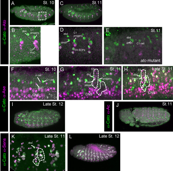
Figure 1.
Cato protein expression in the embryo. Immunohistochemistry with anti-Cato (green) and a second marker antibody (magenta). (A-E) Expression of Cato relative to Ato. (A, B) Stage 10 embryo. Boxed area is magnified in (B). Cato is expressed in the C1 progeny, the dbd precursor and the probable dmd1 precursor; at this stage Ato is mostly in the proneural cluster (PNC) cells for the remaining Ch precursors. (C) Stage 11 embryo. (D) Magnification of two abdominal segments (different embryo to (C)). Cato is now expressed additionally in further Ch SOPs. (E) Stage 11 ato1 mutant embryo, showing that Cato expression remains in the C1, dbd and dmd1 precursors. Note that non-functional Ato protein is still expressed in these embryos. (F-I) Cato expression relative to the SOP marker, Ase. (F) Stage 10 embryo, with Cato in C1. At this early stage Ase is weakly expressed in C1 and A cells, and a glial cell, and strongly expressed in neuroblasts of the CNS. The green channel for the region boxed is shown in the inset image. (G) Stage 11 embryo, showing more ES precursors (expressing Ase) but these do not express Cato at this stage. (H) Late stage 11 embryos, showing that Cato is still not expressed strongly in most ES cells (Ase-positive cells). (I) Late stage 12 embryo, Cato is now expressed widely in PNS cells. (J, K) Cato expression relative to Sens. At early (J) and late (K) stage 12 there is substantial co-expression of Cato and Sens in PNS cells.

