
Figure 1.
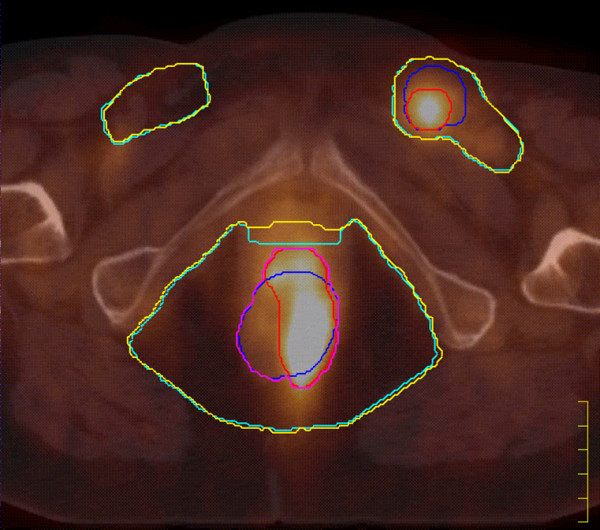
PET/CT image in axial view of a T3N2 case. Different colours are used to highlight the contours of the treatment volumes: CT-GTV (blue), PET-GTV (red), PET/CT-GTV (purple), CT-CTV (light blue), and PET/CT-CTV (yellow). The PET/CT GTV and the PET/CT-CTV were used for treatment purposes.