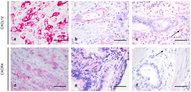
Figure 2. CXCL12 and CXCR4 expression in tumour cells:
Gastric carcinoma samples revealing strong (a) and weak (b) CXCL12 immunoreactivity. Only few cases were CXCL12 negative (c). Note positive CXCL12 staining of blood vessels (arrow). Gastric carcinoma specimens showing a clear cytoplasmic and membranous CXCR4 immunoreactivity were sparse (d). Few samples revealed a weak (e) CXCR4 staining whereas most of the tumours lacked CXCR4 expression (f). Leukocytes served as internal positive control (arrows). Scale bar: a-f: 50 µm.

