
Fig. 4.
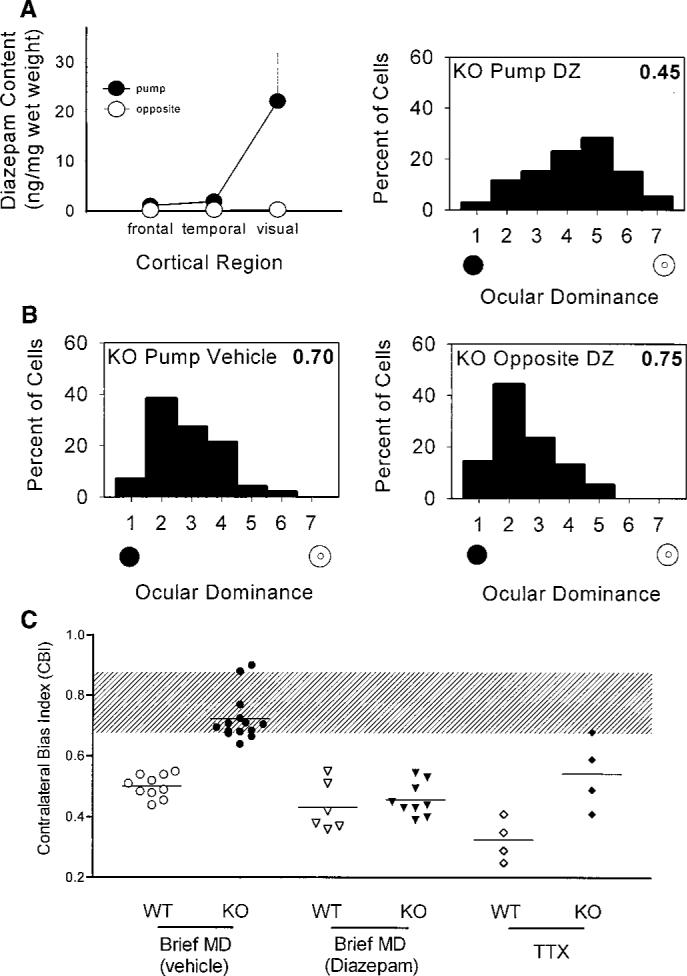
(A) Local diazepam treatment of visual cortex restores plasticity to GAD65 KO mice in vivo. Left panel: Directed infusion restricted diazepam from regions beyond the visual cortex ipsilateral to the cannula (solid circles), as well as throughout the opposite hemisphere (open circles, mean ± SEM, three mice). Right panel: Ocular dominance shifted fully in the KO binocular zone exposed to diazepam concurrent with a brief period of monocular deprivation during the critical period (114 cells, four mice; P < 0.0001, χ2 test vs. nondeprived KO; compare with Fig. 3B). (B) Neither vehicle treatment (left panel, 99 cells, four mice; P < 0.0001, χ2 test vs. diazepam KO above) nor diazepam infusion into the hemisphere opposite the recording site (right panel, 77 cells, three mice; P < 0.0001, χ2 test vs. diazepam KO above) restored the effect of monocular deprivation to GAD65 KO mice. (C) Monocular T TX injections for 4 days during the critical period produced significantly less plasticity in KO than in WT mice (P < 0.05, t test). Untreated and vehicle-treated monocularly deprived (MD) mice are grouped together, as are ventricle- and cortex-infused diazepam-treated animals. Shaded region indicates the range of nondeprived CBIs for both WT and KO mice. Each symbol represents one animal.