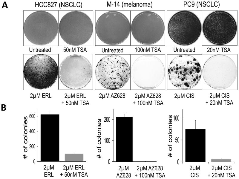
Figure 6. Inhibition of HDAC activity disrupts tolerance to a variety of cancer drugs.
(A) The EGFR-mutant NSCLC cell line NCI-HCC827 was either untreated, treated with TSA (50nM TSA), erlotinib (2μM ERL), or erlotinib plus TSA (2μM ERL + 50nM TSA). After 6 days, the untreated and TSA-treated plates had reached confluence and were Giemsa stained. The erlotinib and erlotinib + TSA-treated plates were similarly fixed and stained after 19 days of treatment. BRAF-mutant M14 melanoma cells were either untreated, treated with TSA (100nM TSA), treated with AZ628 (2μM AZ628), or treated with AZ628 + TSA (2μM AZ628 + 100nM TSA). After 6 days, the untreated and TSA-treated plates had reached confluence and were Giemsa stained. The AZ628 and AZ628 + TSA treated plates were similarly fixed and stained after 39 days of treatment. PC9 cells were either left untreated, treated with TSA (20nM TSA), cisplatin (2μM CIS), or cisplatin plus TSA (2μM CIS + 20nM TSA). After 6 days, the untreated and TSA treated plates had reached confluence and were Giemsa stained. The CIS + TSA treated plates were similarly fixed and stained after 45 days of treatment. Drug treatments were repeated every three days. The experiment was performed in triplicate and representative stained plates are shown.
(B) Quantification of the lower panels in (A) expressed as the number of drug-tolerant colonies observed, with erlotinib-, AZ628- and cisplatin-only treated cells shown in black and the combination of these drugs with TSA shown in grey. Error bars represent the standard deviation from the mean (n=3).

