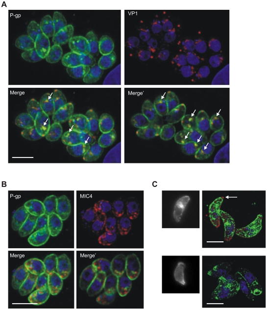
Figure 6. Intracellular localization of T. gondii P-gp.
Confocal microscopy of intracellular T. gondii at 24 h post infection. A. Dual staining using polyclonal mouse anti-P-gp minigene (green) and anti-VP1 (red) antibodies showing partial co-localization of the two proteins (arrows). Nuclear DNA was stained with DAPI. Merge, maximum projection; Merge', single optical section of the deconvolved image stacks. B. Dual staining using anti-P-gp minigene (green) and anti-MIC4 (red) antibodies. Merge, maximum projection; Merge', single optical section of the deconvolved image stacks. C. Wide field fluorescence (left panels) and 3D reconstructed confocal (right panels) micrographs showing differential P-gp distribution in extracellular parasites stained with anti-P-gp minigene antibody (green). The arrow indicates the absence of P-gp staining in the conoid area labeled with anti-T. gondii antibody (red). Nuclear DNA was stained with DAPI (blue). Scale bars: 5 µm.

