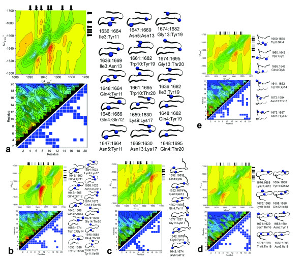
Figure 4.
The structure-spectral correlation for investigated conformations of Beta3s. Letter designations a) native b) Ns c) Cs d) Ch-Curl conformations e) Helix Top Left: The 2DIR plot at Γ of 5 cm-1. Follow vertical axis arrows down until they meet horizontal axis arrows at identified peak. Black arrows highlight current conformation peaks while gray arrows highlight the native conformation peak locations. Bottom Left: The Normal Mode Decomposition plot of residue coupling intensities and structural contact map calculated at 6.7Å cutoff values. Right: The structual representation of the cross peak position and assigned residue. The blue spheres in the structure image represent the residues causing the cross peaks.

