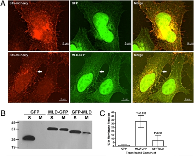
Fig. 2.
RIDVc aa2561-2645 is sufficient to drive GFP to the HeLa cell plasma membrane. (A) HeLa cells transfected with a plasma membrane marker (S15-mCherry) and either GFP or MARTXVc aa2561-2645-GFP (MLD-GFP) were imaged by deconvolution microscopy. Arrow shows overlap at the plasma membrane of mCherry and GFP signals. (B) Representative anti-GFP immunoblot of three separate experiments following membrane fractionation of HeLa cells transfected with the indicated constructs; S, soluble; M, membrane. (C) The average percentage of signal (+/− the standard deviation) in the membrane fraction following fractionation was determined by densitometry of immunoblots for each MLD-GFP construct (n = 3). Tabulated raw densitometry measurements are found in Table S2. A Student's t test was employed to determine the statistical significance of the difference between the amount of GFP versus each GFP-fusion in the membrane fractions; *, significantly different from GFP alone (P < 0.05).

